Gross Anatomy of the Upper Limb
Introduction
Gross anatomy, also known as macroscopic anatomy, refers to the study of the external structures of the body. In the context of the upper limb, gross anatomy encompasses the examination of bones, muscles, tendons, ligaments, nerves, blood vessels, and other visible tissues. This knowledge is crucial for understanding how the upper limb functions and how it relates to overall human physiology.
Overview
The upper limb consists of three main regions: the shoulder girdle, arm (humerus), and forearm. Each region contains various bones, joints, muscles, and other soft tissues that work together to enable movement, support, and sensory perception.
Key Components
- Bones: Provide structural support and protection.
- Muscles: Enable movement through contraction and relaxation.
- Joints: Allow for flexibility and range of motion.
- Nerves: Control muscle function and transmit sensory information.
- Blood Vessels: Supply oxygen and nutrients to tissues.
Detailed Anatomy
Shoulder Girdle

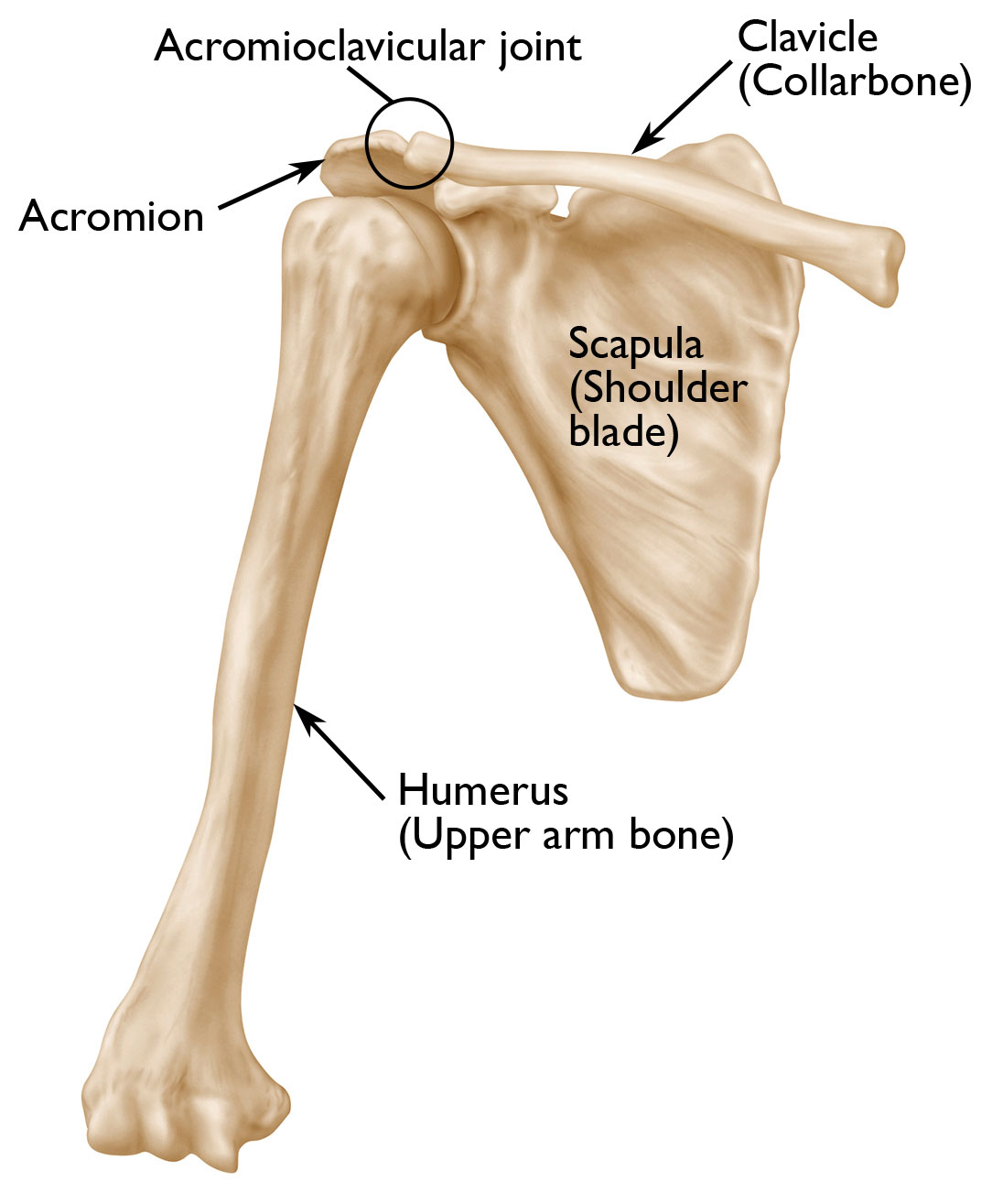
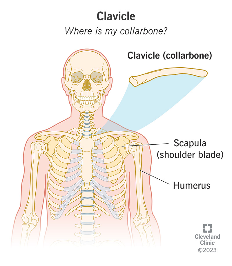
The shoulder girdle or pectoral girdle consists of two clavicles (collarbones) and two scapulae (shoulder blades). It connects the upper limb to the trunk of the body.
The shoulder girdle is the anatomical mechanism that allows for all upper arm and shoulder movement in humans.
Clavicle
- Shape: Long, narrow bone with a conical head.
- Function: Attaches scapula to sternum, supports arm movements.
- Key landmarks: Acromial end, sternal end, costal tuberosity.
Scapula

- Shape: Triangular bone with a flat body and curved edges.
- Function: Provides attachment points for muscles and protects underlying organs.
- Key landmarks: Superior border, inferior angle, acromion process.
Arm (Humerus)
The humerus is the longest bone in the upper limb, extending from the shoulder joint to the elbow.
Humerus Structure
- Proximal end: Head, greater and lesser tubercles.
- Shaft: Diaphysis, epiphyses.
- Distal end: Condyles, trochlea, capitellum.
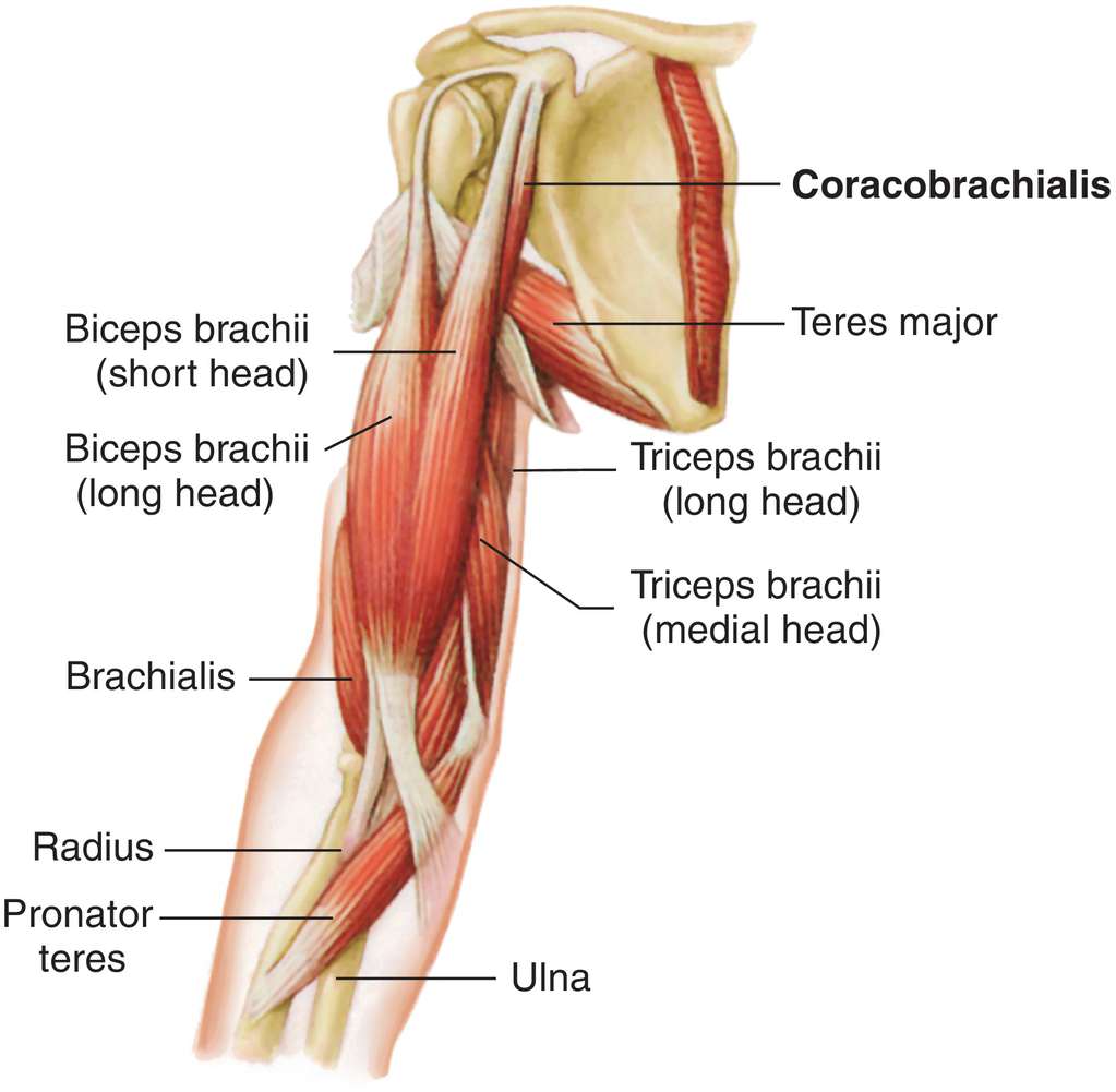
Key Muscles

- Biceps brachii: Flexes elbow, supinates forearm.
- Triceps brachii: Extends elbow.
- Brachialis: Flexes elbow.
- Coracobrachialis: Assists in flexion and adduction of the arm.
Forearm

The forearm contains two long bones: the radius and ulna.
Radius and Ulna
- Radius: Lateral bone, articulates with humerus proximally.
- Ulna: Medial bone, articulates with humerus proximally.
Key Muscles
- Pronator teres: Pronates forearm.
- Supinator: Supinates forearm.
- Flexor carpi radialis: Flexes wrist.
- Extensor carpi radialis brevis: Extends wrist.
Functions and Movements
Understanding the gross anatomy of the upper limb is essential for comprehending its various functions:
- Movement: The upper limb enables a wide range of motions, including flexion, extension, abduction, adduction, rotation, and circumduction.
- Support: The upper limb plays a crucial role in supporting the body during activities like standing, walking, and reaching.
- Sensory Perception: Various receptors in the skin and deeper tissues allow for touch, pressure, temperature, and proprioceptive sensations.
- Manipulation: The hands and fingers are highly specialized for grasping, manipulating objects, and performing fine motor tasks.
- Protection: The upper limb shields vital organs such as the heart and lungs.
Clinical Relevance
Knowledge of upper limb gross anatomy is critical for healthcare professionals, particularly in:
- Diagnosis of musculoskeletal disorders.
- Understanding surgical procedures related to the upper limb.
- Performing physical examinations.
- Interpreting imaging studies.
Examples and Case Studies
Example 1: Radial Nerve Injury
A patient presents with weakness in wrist extension and thumb spreading. Physical examination reveals decreased sensation over the back of the hand and little finger. These symptoms suggest radial nerve damage, likely due to compression at the spiral groove of the humerus.
Example 2: Cubital Tunnel Syndrome
A patient complains of numbness and tingling in the little finger and ring finger when the elbow is flexed. This condition is caused by compression of the ulnar nerve at the cubital tunnel, leading to median nerve dysfunction.
Additional Resources
For further learning and practice, consider exploring:
- Anatomical models and dissection guides.
- Interactive online tutorials.
- Cadaveric dissections under supervision.
- Radiological imaging studies (X-ray, CT, MRI).
- Clinical case studies and literature reviews.
By mastering the gross anatomy of the upper limb, students gain a fundamental understanding of human structure and function. This knowledge serves as a foundation for more advanced studies in fields such as medicine, surgery, physiotherapy, and biomechanics.
Remember, practical application of anatomical knowledge is key. Regular practice with cadavers, prosections, or virtual reality tools can significantly enhance your understanding and retention of this material.
As you continue your journey in studying gross anatomy, keep in mind that each component of the upper limb works in conjunction with others to form a complex system. A thorough comprehension of these relationships will greatly benefit your future studies and professional practice.
Definitions of Medical Terms
- Acromion: A bony process on the scapula that forms the highest point of the shoulder.
- Adduction: Movement toward the midline of the body.
- Biceps brachii: A muscle of the upper arm responsible for flexing the elbow and supinating the forearm.
- Capitellum: A round knob on the distal humerus that articulates with the head of the radius.
- Circumduction: Circular movement of a body part, such as the arm, involving flexion, extension, abduction, and adduction.
- Coracobrachialis: A small muscle in the front of the arm that helps in flexion and adduction of the arm.
- Cubital Tunnel: A passageway on the inner side of the elbow, where the ulnar nerve runs through.
- Diaphysis: The shaft or central part of a long bone.
- Epiphysis: The rounded end of a long bone, which is involved in forming joints.
- Flexion: Bending a joint to decrease the angle between two bones or body parts.
- Greater and lesser tubercles: Small, rounded projections on the proximal end of the humerus, where muscles attach.
- Pronation: Rotational movement where the palm or forearm faces downward.
- Proximal: Closer to the point of origin or attachment.
- Radius: One of the two bones of the forearm, located on the lateral side (thumb side).
- Radial nerve: A major nerve in the arm responsible for controlling muscles that extend the wrist and fingers.
- Supination: Rotational movement where the palm or forearm faces upward.
- Trochlea: A pulley-shaped structure on the humerus that articulates with the ulna.
- Ulna: One of the two bones of the forearm, located on the medial side (pinky side).
- Ulnar nerve: A major nerve running along the inner side of the arm and forearm, often associated with the "funny bone" sensation.
